GW researchers fabricated a vascularized patch of cardiac tissue suitable for cardiac regeneration using 3D bioprinting. Cardiovascular disease involving myocardial infarction is a major cause of morbidity and mortality worldwide. Following myocardial infarction, adult cardiac tissue cannot significantly repair or regenerate itself. Current therapies include autografts, allografts, xenografts, and artificial prostheses. Disadvantages of the standard of care include donor tissue shortages, immune rejection, required anticoagulation therapy, and limited durability. 3D bioprinting avoids or mitigates these disadvantages.
Competing 3D printed cardiac patches are very thin and have simple vascularization patterns. GW’s thick “sandwich” patch has a layer of vessels printed between two layers of muscle tissue. The researchers combined two types of 3d bioprinting to create the thick biomimetic patch. Stereolithography-printed anisotropic honeycomb fibers laden with cardiomyocytes mimic heart muscle and extrusion-printed human umbilical vein endothelial cell laden vessels provide blood flow required to support thick tissues. After printing, perfusion culturing enhances the development of the engineered tissues. In vitro and in vivo (mouse) studies reveal excellent cardiomyogenesis and angiogenesis. The anisotropic fibers provide structural support in preventing the heart wall from dilating, and the complex perfusable vasculature with the fibers aid in regeneration of the cardiac tissue. Studies with thicker patches in larger mammals are ongoing.
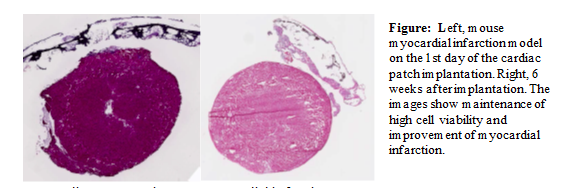
6 weeks after implantation.This image shows maintenance of high cell viability and improvement of myocardial infarction.
Applications:
- Cardiac regeneration post myocardial infarction
Advantages:
- Anisotropic fibers support direction dependent contraction
- Anisotropic fibers prevent dilation of heart wall
- Perfusable complex vasculature enables thick patches (1cm performed)
- 3D bioprinting parameters can be adjusted to fit patient-specific requirements