GW researchers generated a vascularized and highly osteogenic bone construct with a biphasic structure using a multiple 3D printing platform. Traditional tissue engineering techniques face major obstacles in creating multiple tissue structures and vascularization of tissues. 3D bioprinting allows for control over the location of cells and biomaterials; however monotypic model printing has limitations in creating multi-structured tissues. Vascularization is essential to prevent cell death in internal regions of large implants.
Our solution, dual 3D bioprinting, utilizes fused deposition modeling (FDM) and stereolithography (SLA). The dual platform creates a bone-mimicking construct with a soft organic matrix surrounding a hard mineral structure. The construct has a network of large vessels and smaller capillaries. This is done by alternate deposition of biodegradable polylactic acid (PLA) fibers and cell-laden gelatin methacrylate (GelMA) hydrogels. Bone Morphogenic Protein 2 (BMP2) and vascular endothelial growth factor (VEGF) are regionally immobilized onto the scaffolds to promote osteogenesis and angiogenesis. This served as a proof of concept and future research will expand the multiple 3D printing platform to other types of tissue regeneration.

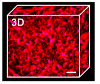
Figure: Vascular capillary network.
Applications:
- Manufacturing 3D printed vascularized bone implants
Advantages:
- Regional immobilization of bioactive factors
- Suitable for large bone implants