When diagnosing developmental neurological disorders, physicians start by labeling (parcellating) brain regions. Growth abnormalities in specific regions correspond to deficits in particular neurological functions. Historically, cortical parcellation is done manually by trained experts, which is costly and time-consuming (1-5 days for a single brain scan from magnetic resonance imaging).
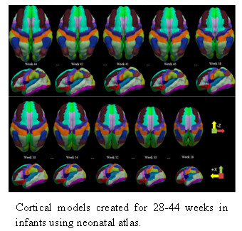
Researchers at GW devised a novel algorithm to automate parcellation of the neonatal brain at different stages of prenatal and neonatal development. The method starts with a manually labelled gestational week 41 atlas (average of multiple individuals) and applies the labels to an atlas of brain scans from various prenatal and neonatal developmental stages (gestational weeks 28-44).
The resulting model can be used as a diagnostic aid in accurately determining the presence of deformities in the cortical surfaces of the neonatal brain.
Applications:
- Detect neurological disorders in premature infants, neonates, and aging patients
- Label and track cortical regions and their growth
- Compare a developing or aging brain at two time points
- Compare two different brains at a single timepoint
Advantages:
- Inter-subject variability in cortical geometric patterns no longer poses a problem
- Can be extrapolated to any stage of gestational development
- Multiple neurological disorders are diagnosable