The minimally invasive MitoScope assesses the metabolic rate of heart tissue in vivo, non-destructively, and in real-time. MitoScope measures the mitochondrial enzyme activity of the heart to provide unique insight that could improve the treatment of heart failure, coronary heart disease, diabetes, and many other diseases that alter myocardial metabolism. Existing technologies for metabolic assessment of the heart require biopsies and subsequent in vitro lab analysis. The MitoScope circumvents these limitations to provide real-time information.
MitoScope is the first in vivo application of the metabolic rate measurement approach called Enzyme-Dependent Fluorescence Recovery after Photobleaching (ED-FRAP). ED-FRAP quantitatively measures the distribution of enzymatic activity within small samples or cells. The method relies on the selective destruction of the metabolite, NADH, using a photo-bleaching ultraviolet light pulse and measuring the rate of enzymatically driven recovery of the metabolite. The rate of fluorescence recovery indicates the rate of mitochondrial NADH production and therefore the capacity of living cells to produce energy.
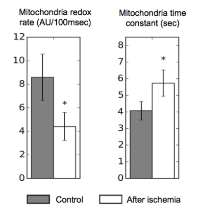
GW researchers developed MitoScope to apply ED-FRAP to living contracting hearts using light-emitting diode (LED) technology for non-destructive, repeatable measurements of cellular NADH production. Experiments on perfused living hearts demonstrated, for the first time, reduced ability of cardiac cells to produce energy after ischemia-reperfusion injury, an outcome typically encountered during a heart attack and a common complication of heart failure.

Figure: Mitochondrial energy production rate of the left ventricle of the heart.
Applications:
- Repeatable measurement of the cellular energy production rate of heart tissue and other metabolically active tissues
Advantages:
- Non-destructive
- Minimally invasive
- Real time results